Submission ID: 011275
Title:
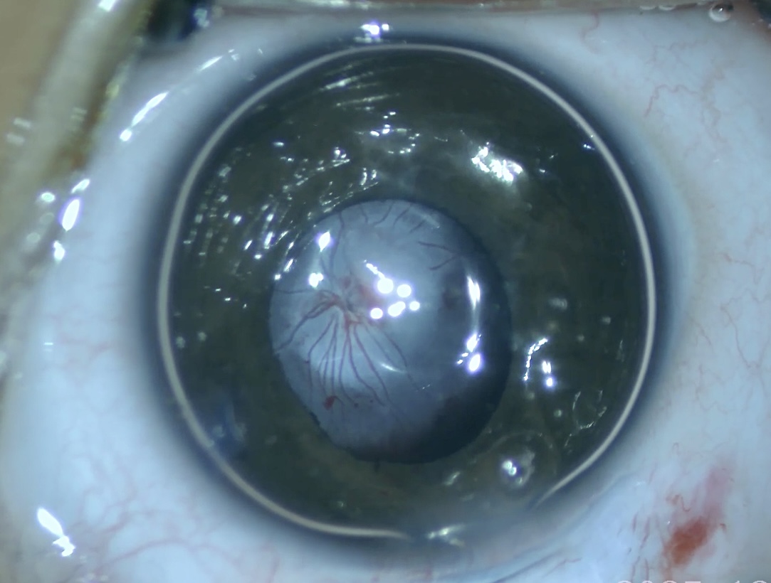
The Eye That Stayed Foetal
Category:
I haven´t seen that before
Description:
This intraoperative image reveals persistent tunica vasculosa lentis and retrolental fibrovascular tissue, demonstrating preserved fetal vasculature. Captured at 45 days of life, coaxial microscope illumination sharply delineates the embryologic circulation and associated tractional distortion of the lens. The photograph offers a rare glimpse of ocular development arrested in vivo, illustrating how failed regression of primary vitreous architecture can influence visual outcomes.
Frozen foetal circulation
Operating microscope photography
IMG_6069.jpeg
Go Back