Submission ID: 010855
Title:
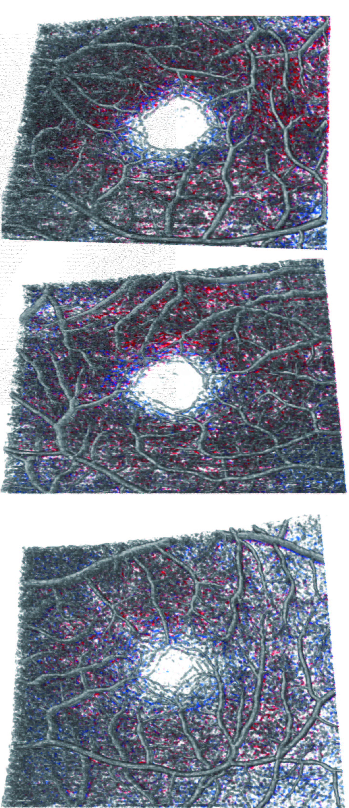
three-dimensional visualization of vessel density across the full-thickness retina
Category:
OCT, OCT-A
Description:
with SVP in gray, ICP in blue, and DCP in red.Top row: amblyopic eye (OS);Middle row : fellow eye in anisometropic amblyopia patient (OD); Bottom row: healthy control (OS).Vessel density is highest in healthy controls, followed by fellow eye in anisometropic amblyopia patients, and lowest in amblyopic eyes.First, OCT and OCTA data are fed into a multitask convolutional neural network(CNN )to segment 3D RVs from volumetric OCT/OCTA data directly.Second, based on the 3D segmentation results, the vascular network is reconstructed in 3D, and a vessel skeleton map is extracted. Third,3D metrics are extracted to analyze the retinal vasculature quantitatively, including vessel volume density (VVD) ,which is calculated as the ratio between retinal vessels to the total retinal volume within the measurement region.
three-dimensional visualization of vessel density across the full-thickness retina
Spectralis II OCTA (Heidelberg Engineering GmbH, Heidelberg, Germany)
3DOCTA.jpg
Go Back