Submission ID: 010850
Title:
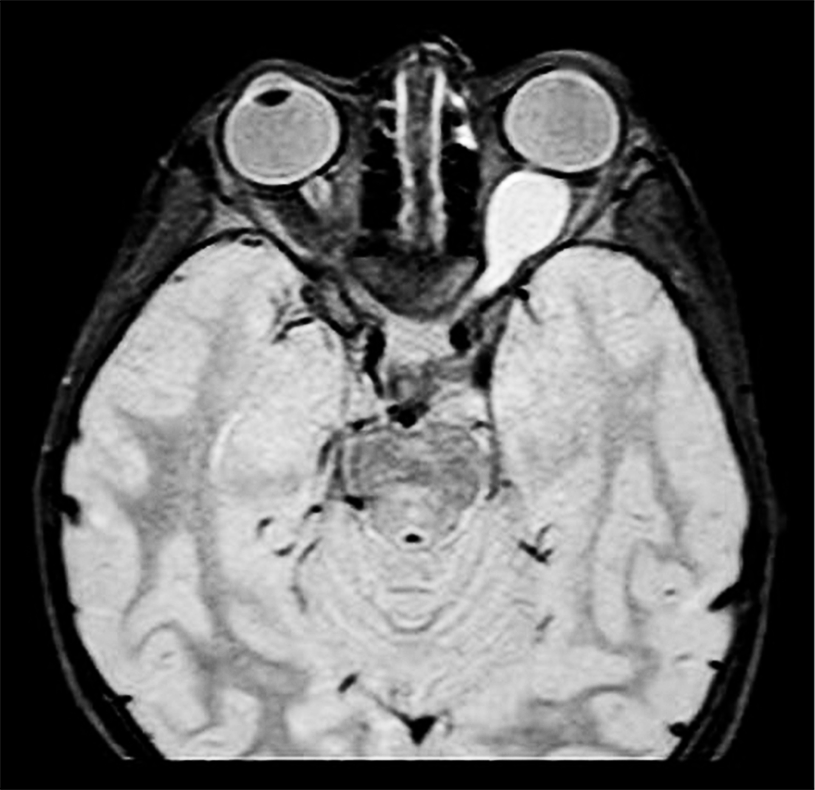
The Silent Intruder: Optic Nerve Glioma in Neurofibromatosis Type 1
Category:
I haven´t seen that before
Description:
Axial MRI (T1-weighted) showing fusiform enlargement of the optic nerve–glioma characteristic of Neurofibromatosis Type 1 (NF1). The involved optic nerve appears thickened with a homogeneous signal pattern, producing the classic “pseudo–cerebriform” contour. The surrounding orbital fat planes are preserved, while the contralateral optic nerve maintains normal caliber. This imaging elegantly illustrates the hallmark benign, slow-growing nature of NF1-associated optic pathway gliomas, often presenting in childhood with insidious visual decline, proptosis, or strabismus. The bilateral symmetry of the globes contrasted against the distorted optic nerve architecture makes this a striking radiologic example.
The Silent Intruder: Optic Nerve Glioma in Neurofibromatosis Type 1
High-resolution MRI Orbit Protocol using 1.5T Siemens Magnetom System with axial T1-weighted sequences
NF.jpg
Go Back