Submission ID: 010813
Title:
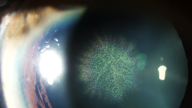
Burst of colours- Sunburst/sun flower cataract in Wilsons disease
Category:
Anterior Segment photography
Description:
Wilson’s disease(WD) is a rare autosomal recessive disorder causing decreased hepatic copper
excretion and accumulation of copper in various tissues like brain, liver, kidney, cornea. Typical
ophthalmological manifestations include Kayser-Fleischer ring (KFR)- extracellular deposition of
copper in Descemet’s membrane and sunflower/sunburst cataracts - copper deposition in anterior
and posterior capsule .A 27-year-old presented with a rare combination of bilateral KF rings and cataract with central green disc and radiating spokes resembling sunflower.Sunflower cataract is a rare manifestation where as KF rings are seen in 97% of WD with neurological signs.
Burst of colours- Sunburst/sun flower cataract in Wilsons disease
slit lamp photography
wilsons lens.jpg
Go Back