Submission ID: 010764
Title:
Wonder drug turns foe
Category:
External photography
Description:
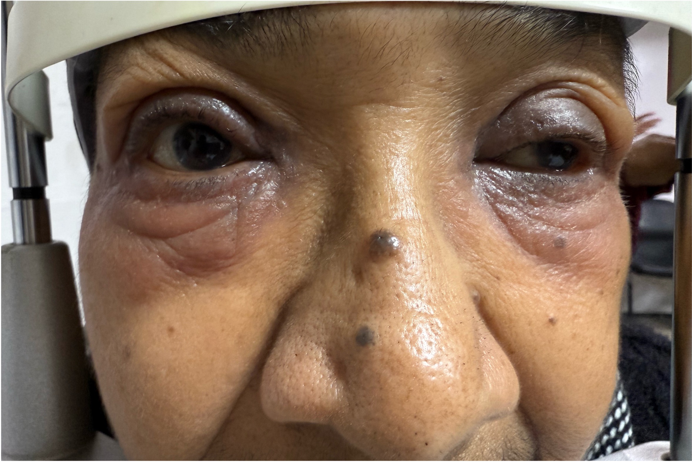
Photograph of a patient who developed severe periocular contact dermatitis, 4weeks after using topical dorzolamide. Periocular involvement is bilateral, eczematous swelling of the lids (lid edema), redness alongwith itching/pruritis. Allergic contact dermatitis to topical dorzolamide is usually delayed (range roughly 4–40 weeks) after initiation of topical dorzolamide, localized reaction and does not necessarily predict a systemic reaction to oral carbonic anhydrase inhibitors (CAIs)
Wonder drug turns foe
Phone camera
1.jpg
Go Back