Submission ID: 010603
Title:
Temporal CHRPE Spotlight: Healthy Disc, Fresh Reflex & Retina On
Category:
Posterior Segment photography
Description:
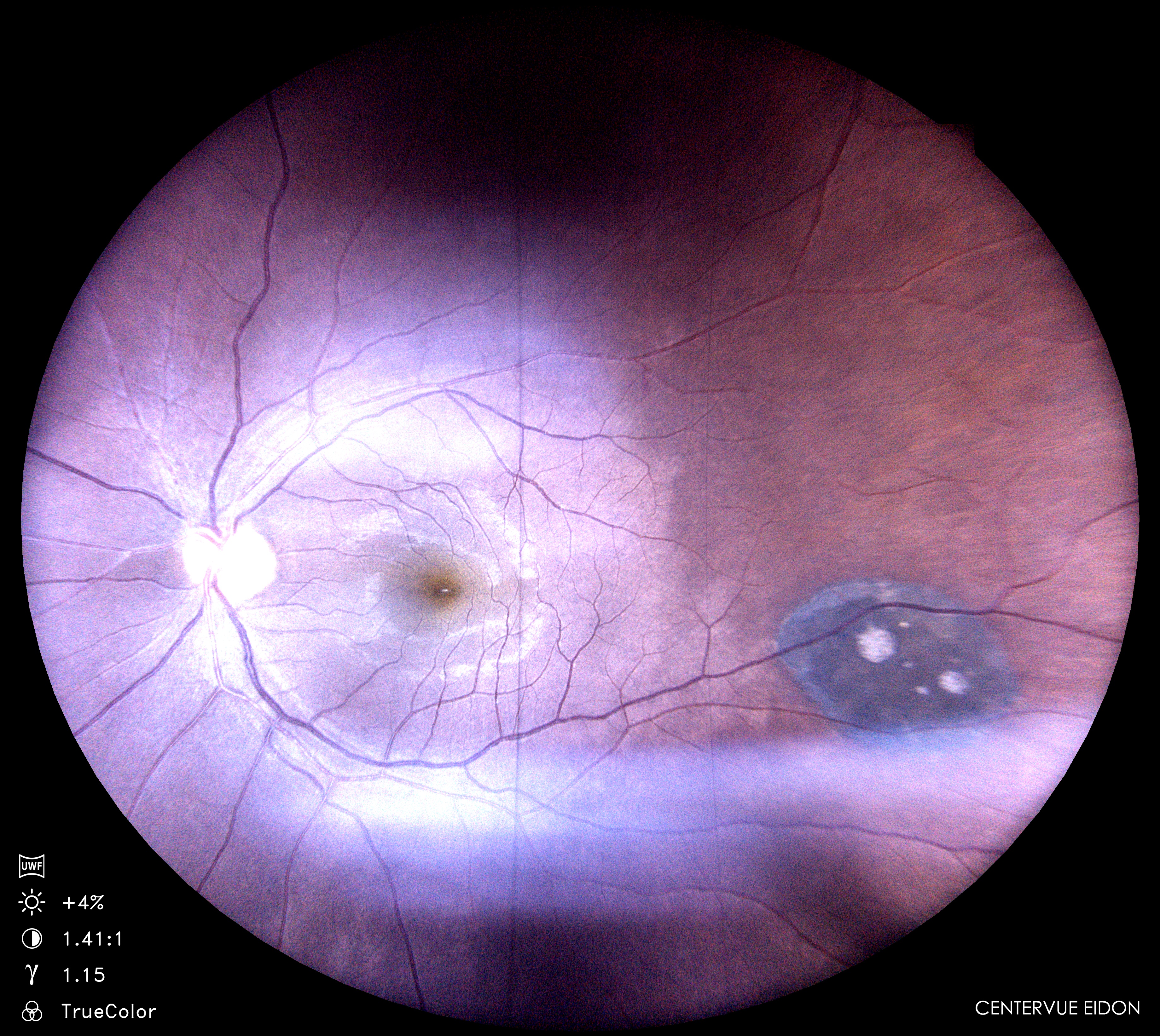
The fundus image shows a fully attached retina with a bright foveal reflex, indicating a healthy macular contour. The optic disc appears normal with well-defined margins and physiologic vessel pattern. A solitary congenital hypertrophy of the retinal pigment epithelium (CHRPE) lesion is seen in the temporal quadrant, presenting as a flat, well-circumscribed dark patch with uniform pigmentation
Temporal CHRPE Spotlight: Healthy Disc, Fresh Reflex & Retina On
Eidon
3.jpg
Go Back