Submission ID: 010062
Title:
The Fiery Glow - Carotid Cavernous Fistula
Category:
External photography
Description:
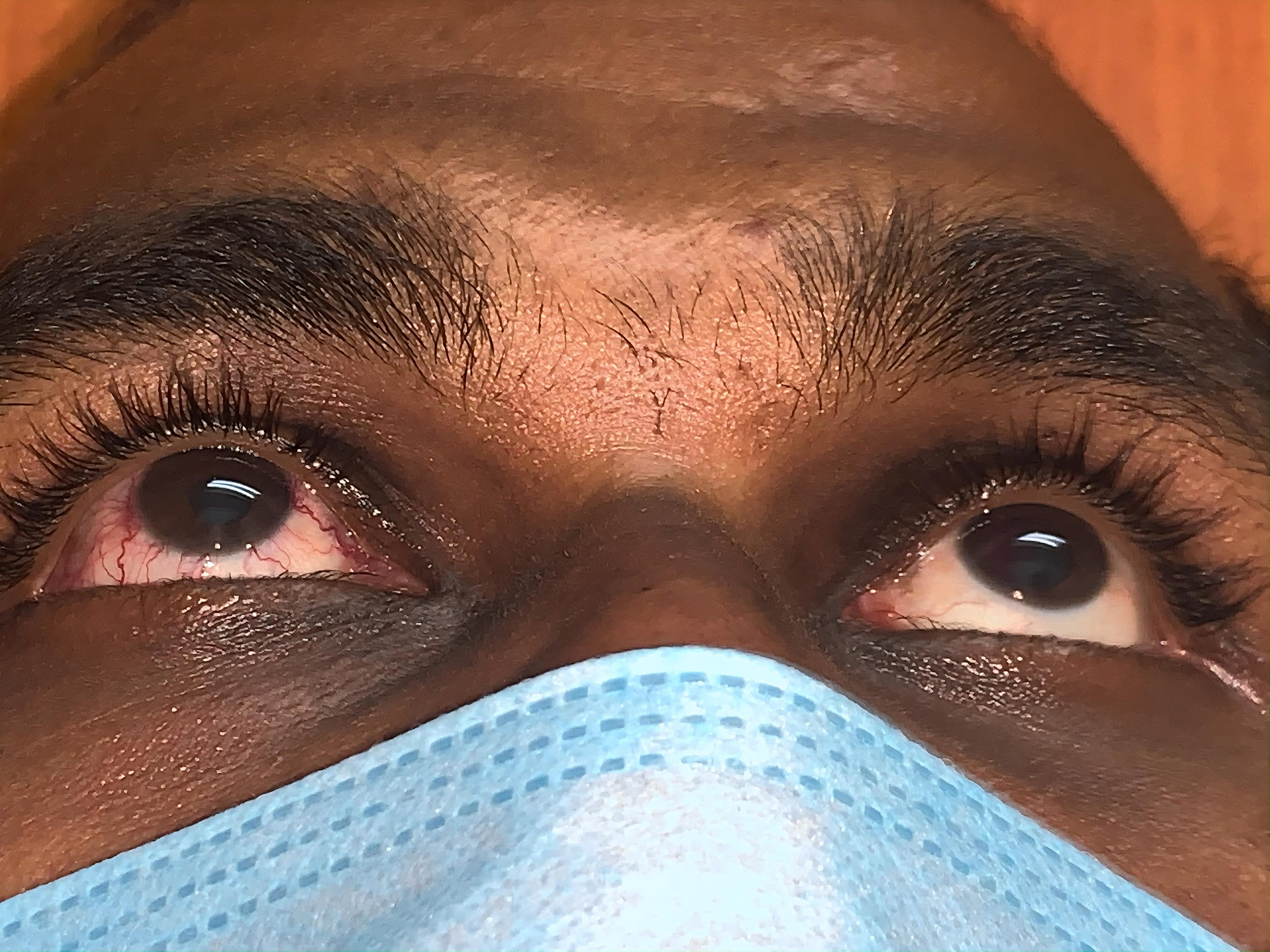
This clinical photograph shows dilated episcleral vessels in the right eye and normal study in left eye. Based on history and imaging studies, carotid cavernous fistula (CCF) was diagnosed. The conjunctival hyperemia associated with CCFs is a result of the arterialization of conjunctival and episcleral veins, consisting of distinct tortuous corkscrew blood vessels that converge at the limbus.
The Fiery Glow - Carotid Cavernous Fistula
iPhone 13 Pro
010062.jpeg
Go Back