Submission ID: 010046
Title:
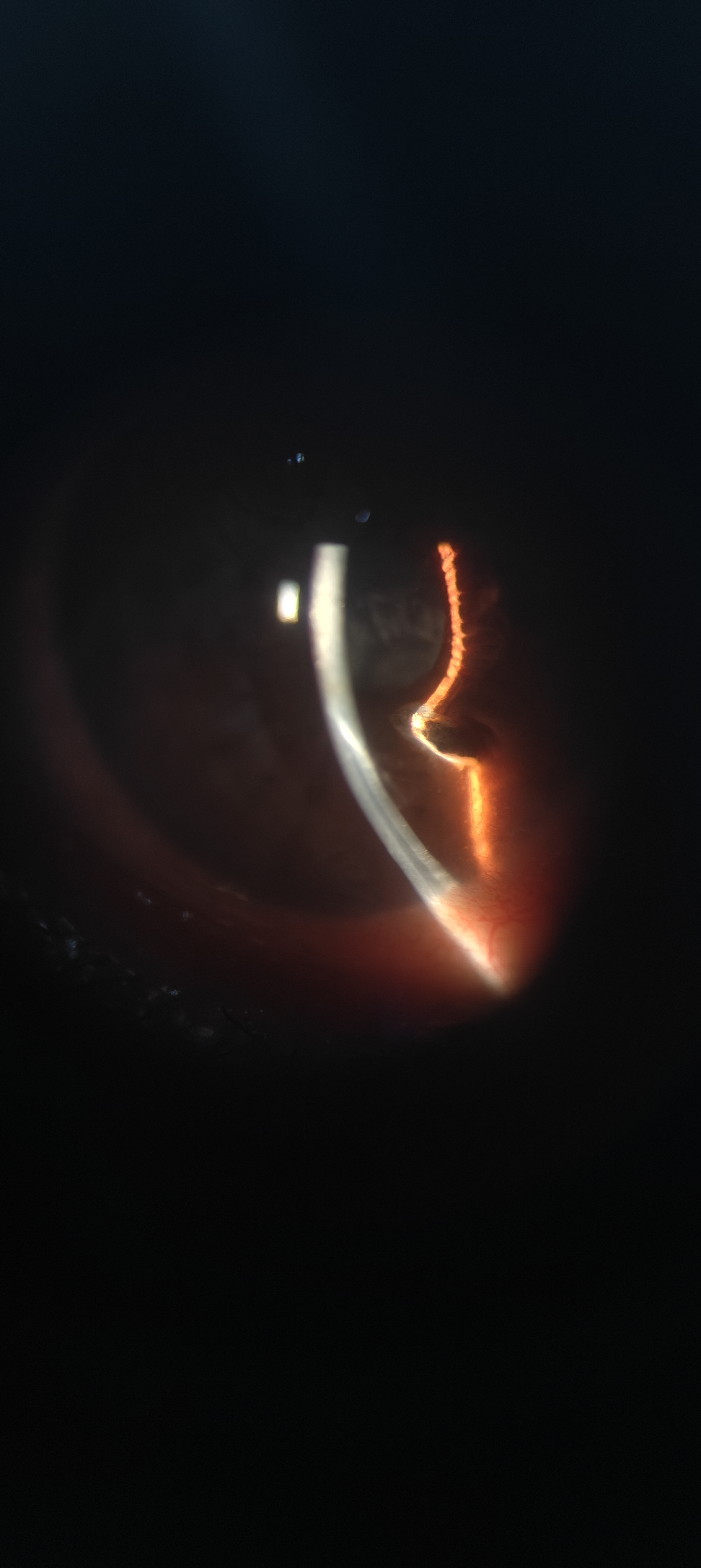
The Foveal Contour Illusion: A Curious Reflection from an Iris Foreign Body
Category:
Arts and Crafts
Description:
A metallic foreign body embedded in the iris for four months presents an extraordinary optical illusion—the slit beam reflection tracing a perfect foveal contour within the anterior chamber. What seems like a macular OCT image is, in fact, the eye’s own artistry, sculpted by light, reflection, and trauma. This photograph exemplifies how science and art coexist within ophthalmology, transforming pathology into visual poetry. Beyond its clinical message of vigilance in detecting retained intraocular foreign bodies, it reminds us that every image in medicine can hold a story—where precision meets perception, and art quietly resides within science.
Go Back
